

The topic of this blog is the lateral cuneiform bone. You probably have never heard of this bone. There are many bones in the human foot, 26 total. Names of foot bones that may be more familiar to you are the metatarsal bones, phalanges, calcaneus (heel bone), and talus (ankle bone). You may have heard of these bones because pain or fractures may occur in their location. The lateral cuneiform bone has a significant role in foot function, however it does its job silently. It does the work it was built to do, and you’ll seldom hear from it as it’s very rare to get injured. It’s part of a group of bones called the tarsal bones that we’ll discuss before diving into the lateral cuneiform bone specifically.
Basic foot anatomy
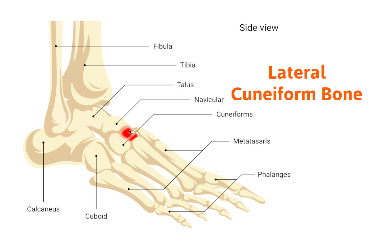
The foot can be separated into three separate areas. The rearfoot, is comprised of the calcaneus (heel bone), and the talus (ankle bone). The forefoot is composed of the metatarsal bones and phalanges and finally, the area in between is called the tarsal bones.
The tarsal bones. There are five bones between the rearfoot bones and the forefoot bones. Together they form the transverse arch of the foot that supports the arch. Starting towards the ankle there is the navicular bone, on the medial side of the foot, which forms a joint with the talus bone (ankle bone), and the cuboid bone which forms a joint with the calcaneus (heel bone). On the other side of the cuboid bone towards the forefoot are the fourth and fifth metatarsals that articulate with the cuboid bone forming separate joints. The cuboid and fourth and fifth metatarsals form the lateral arch. The navicular bone as we move away from the ankle towards the forefoot has three bones that articulate with the navicular to the first, second, and third metatarsals. These wedge-shaped bones are referred to as the first, second, and third cuneiforms. The medial cuneiform bone is the largest with the second being the smallest. They are also called the medial cuneiform, intermediate cuneifor,m and lateral cuneiforms.
The lateral cuneiform referred to as the external cuneiform,(middle cuneiform) which is the topic of this blog, forms the joint with the navicular bone and also the middle cuneiform medially and lies medial to the cuboid bone. It is also directly connected to the third metatarsal and does have some articulation with the second and fourth cuneiforms sharing articulation with the second and fourth metatarsals. It directly articulates with four bones. Together the four bones we just discussed form the medial arch. This arch compared to the lateral arch has more strength and can withstand more weight.
One of the functions of the lateral cuneiform bone is to serve as an insertion of the tibialis posterior tendon. The posterior tibial tendon is a very important tendon for normal foot function. It’s primary function is the support of the arch and decelerator pronation which means flattening of the foot. With part of the posterior tendons inserted into the foot on the bottom of the third cuneiform, it has an important function to stabilize the arch. Without this tendon, the foot loses much of the support in the arch. It also serves as the insertion of the flexor hallucis brevis muscle which assists in flexing the great toe downward.
Injuries to the lateral cuneiform bone
These are very rare. As I mentioned earlier it silently does its job assisting in supporting your arch. However, there is one type of injury called a Lis franc’s joint injury. Lis Franc’s joint refers to the joints formed by the metatarsal bones and tarsal bones. This injury is caused by a mechanism where the forefoot bones are forced downward in relationship to the tarsal bones. This may happen when landing on the ball of the foot forcing the ankle joint to move downward. The foot is not built well to withstand this type of force and as a result, you can suffer from multiple injuries to the bones including fractures and dislocations. This injury involves the bones and joints that form Lis Franc’s joint. In this situation, there may be an injury to the third cuneiform and its joints.
So don’t get too alarmed if you’re getting pain in your midfoot area as its most likely not from this bone. If needed the specialist may offer x-rays, MRI, or CAT scan to rule it out. However, I hope this blog gave you a better concept of the complexity of the human foot and how well if performs for us on a daily basis. You now understand the importance of the tarsal bones and more specifically the lateral cuneiform.
If you’re experiencing persistent midfoot pain or any other foot-related issues, it’s important to seek professional care. At Anderson Podiatry Center, our specialists are dedicated to diagnosing and treating foot conditions with expertise and compassion. Whether you’re in Broomfield or Fort Collins, our team is here to help you get back on your feet. Don’t hesitate to schedule an appointment today and let us take care of your foot health.
Call us today at our Fort Collins location (970) 329-8158, Broomfield location (303) 997-2795, Surgery Center (970) 329-8158, or use our online scheduling system to book your appointment.