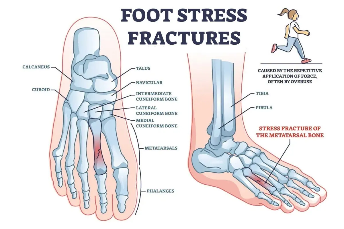
Stress fractures are one of the common athletic injuries in the foot and ankle. It has been said that they may comprise 20% of these injuries. A stress fracture is caused by putting too much stress through a bone that does not have time to recover. At first, it may be considered a bone bruise but with repetitive stress, a break may develop in the bone. By definition, a stress fracture is not typically from one traumatic episode. The challenge of a stress fracture in the foot is that the diagnosis could be missed, and treatment could be delayed. The message of this blog is that the earlier a stress fracture is detected, the faster you’ll receive treatment. Thus providing you with the opportunity to get back to regular activity faster.
Symptoms of a Stress Fracture: How do you know if you have a fracture in your foot?
Generally, no matter where a stress fracture is occurring there tends to be swelling around the injured bone. Typically there will be a high amount of pinpoint pain directly in that area. Usually pressing directly on the bone will cause pain. The pain associated with the stress fracture may make walking difficult!
Just because you can walk on your foot does not mean you don’t have a fracture.
The most common location for stress fractures in the foot tends to be the metatarsal bones. These are the long bones that start in the mid-foot area and extend down to the toes. Ending with the weight-bearing bones of the ball of your foot.
Risk Factors: How Does a Stress Fracture Occur?
High Impact Activities
High-impact activities are common causes of stress fractures. These could include sports such as track and field, running, or other high-impact sports such as tennis, basketball, and football. All of these are repetitive activities that put added stress on the foot and ankle which can lead to a stress fracture.
Improper training can lead to stress fractures. Doing too much too soon or not having rest days between the high-impact activities will not allow your body time to repair. It is important to have rest days for the body to rest or you may develop stress fractures.
Foot Types
Some foot types are more prone to stress fractures. Some people are born with a shorter 1st metatarsal, and this is the bone that makes up the great toe joint. If this bone is excessively short, this will cause more weight bearing on the 2nd and 3rd metatarsal bones.
Bone Insufficiency
Fractures are more common with the following:
Hyperparathyroidism
If the parathyroid gland is hyperactive, the parathyroid hormone will take calcium from the bone and put it in the bloodstream thereby creating weakness in the bones.
Osteoporosis
Osteoporosis appears more frequently in women than in men and it could be related to eating disorders or menstrual dysfunction.
Vitamin D
Vitamin D deficiency can also cause weakness of bone and can be more common in winter months when sun exposure is more limited.
Diagnosing a Stress Fracture
When you see a foot specialist, they will examine your foot and look for swelling and pain. The clinical exam itself is important and will lead the podiatrist to suspect a fracture.
X-rays
X-rays are taken but stress fractures may not be seen if taken soon after the symptoms have started. Oftentimes, the fracture is so small that it is not seen on X-rays. Ten days after the fracture starts, new bone will form that is visible on x-ray and this is called a bone callus. That is why X-rays may show no signs of a stress fracture but if the x-ray is repeated ten days later, the bone callus will be seen, and the diagnosis can be made.
MRI – Magnetic Resonance Imaging MRI
This can be considered the gold standard and is highly recommended especially if x-rays are negative for a stress fracture, but the symptoms correlate to a stress fracture.
Bone Scan
A bone scan can also be performed which will more accurately detect a stress fracture than x-rays.
Treatment
With very few exceptions, the use of a CAM walker is recommended. For many people, they can continue weight bearing in a CAM walker. The use of a CAM walker can last from 3 to 6 weeks depending on the severity of the stress fracture. In the rare instance that the bone is displaced at the fracture site, this may necessitate the need for non-weight bearing before going to weight bearing in the CAM boot. The reason for this is to prevent further displacement of the bone.

Location of Stress Fractures in the Foot
There are many types of stress fractures.
Metatarsal bones
These are the most common bones to have stress fractures especially the 2nd and 3rd metatarsals and to a lesser degree the 4th and 5th metatarsals.
Heel Bone: Calcaneus
This is an uncommon stress fracture. When a stress fracture occurs in the heel, it is usually quick to heal because it has a very good supply.
Tarsal bones
There are many cube-shaped bones in the midfoot area where stress fractures may also occur but again are not as frequent as the metatarsal bones.
Sesamoid bones
Two small bones underneath the big toe joint function much like the patella in the knee joint. These occasionally can fracture and can be caused by trauma to the big toe area but less frequently, can be caused by overuse.
In summary, early detection of a stress fracture is vital. So, it is important not to ignore your symptoms, especially if they’re located in the region of the metatarsal bones as early detection is important. It can reduce the time to heal the stress fracture if early treatment is administered.
Call us today at our Fort Collins location (970) 329-8158, Broomfield location (303) 997-2795, Surgery Center (970) 329-8158, or use our online scheduling system to book your appointment.


