
Freiberg’s disease is a rare presentation in the foot that can be misdiagnosed and overlooked. To better understand what it is we first need to go over the basics of how your bones develop in your foot.
What are the symptoms of Freiberg’s disease?
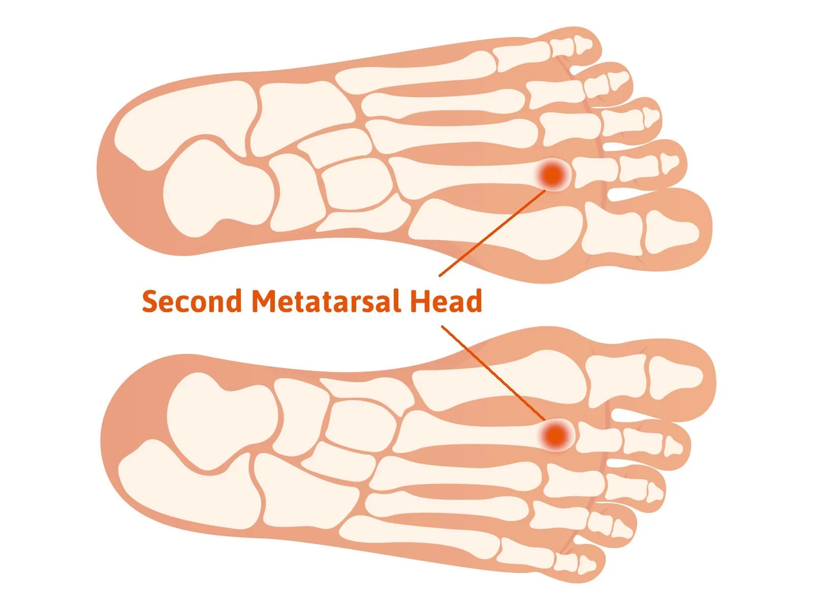
The patient will have pain in the ball of the first. The area that is affected is the metatarsal head. Pain is most common at the base of the second metatarsal head. and rarely at the 4th or 5th toe. There may be swelling at this joint. There may also be a limited range of motion compared to the adjacent joint. Rolling up on the ball of the foot when walking may create pain in this area.
The basics of growth plates in the foot
You may have heard of a term called growth plates. We all have them as our bones develop in childhood. A growth plate is in the long bones of our body and it’s from this region the bone develops to make a short bone longer. The growth plate is comprised of cartilage. If you look at an x-ray of a child’s foot you see a dark area going across the bone which contrasts with the white appearance of the bone. Cartilage does not show up on x-ray and that explains the dark area. For a bone to get longer the cartilage cells in the growth plate area multiply and then transform into bone cells and over time the bone will elongate to its adult length. Then at puberty the growth plates will close, meaning the cartilage that was once there will transform into bone. The growth plate will no longer be seen on an x-ray.
What causes Freiberg’s disease?
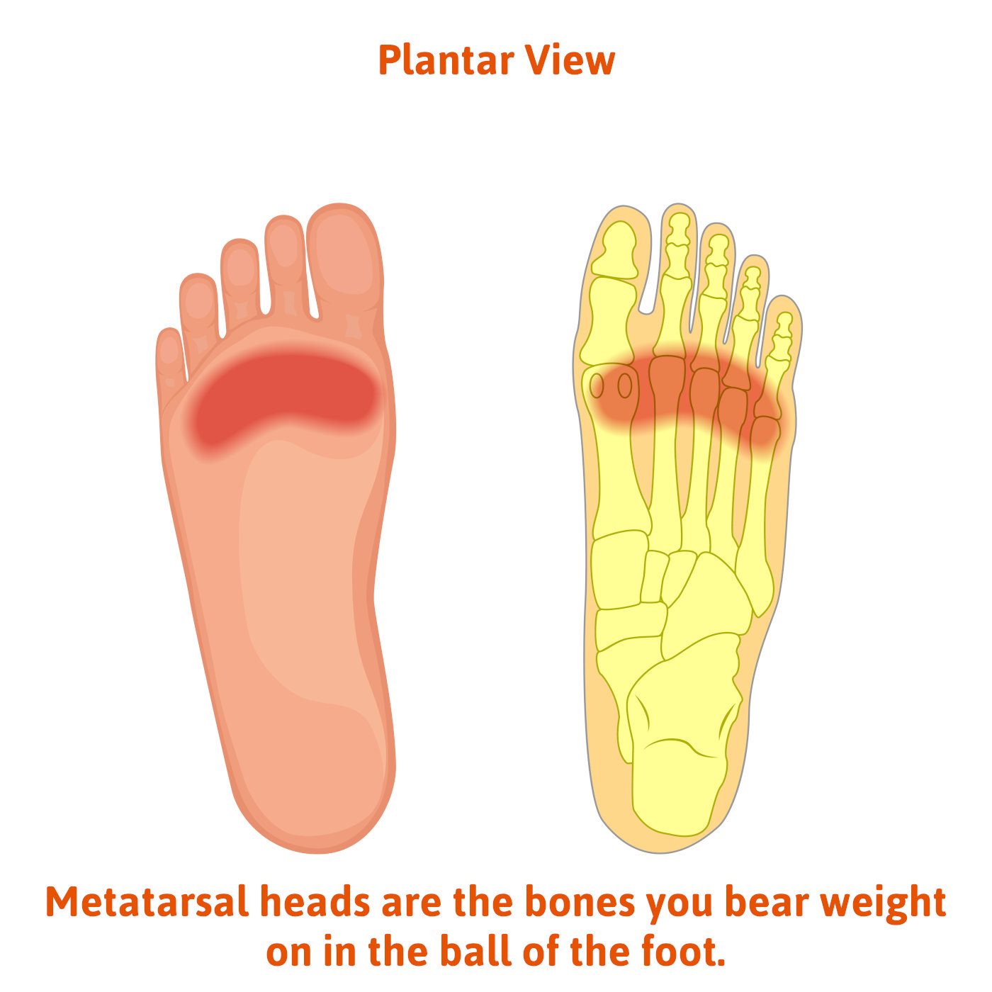
When Frieberg’s disease occurs the growth plate has been damaged. With this damage the blood supply to the growth plate may occur. This will then create what is referred to as avascular necrosis to the metatarsal head which is the area where the growth plate is located. The metatarsal bones are the long bones that goes from the midfoot area down to the ball of the foot. Metatarsal heads are the bones you bear weight on in the ball of the foot. The national library of medicine has labelled it as “an uncommon yet clinically significant condition“.
As the word implies, blood supply is diminished (avascular) and the bone will die away (necrosis). With diminished bone directly beneath the cartilage of the metatarsal head the cartilage deteriorates. This cartilage damage will cause them to become arthritic. There are two primary reasons this may happen.
- Trauma to the area in childhood. The story that I will commonly hear is that I got stepped on, many times by a horse, but any type of direct blow to this area could damage the growth plate that then goes undetected.
- Overload on the metatarsal head – if the metatarsal is longer in relation to the neighboring metatarsals the stress in this area as the bone is developing can also be a cause. This is especially true in the second metatarsal. It may also be from wearing high heels that would overload the second metatarsal.
What is done to diagnose the problem of Frieberg’s disease?
- clinically there will be pain at the metatarsal phangeal joint involved. The motion may be limited and painful to move.
- X-rays will show evidence that the joint space may be narrower, and the metatarsal head may appear flatter than normal.
- MRI- An MRI can more specifically evaluate the joint and reduction of blood supply to the area.
Treatments for Freiberg’s disease
Conservative treatment
- Immobilization – resting the area by using a Cam walker for 4-6 weeks (about 1 and a half months) may be helpful. However, this is unlikely to give long-term relief.
- Orthotics – The use of custom-made inserts may help to reduce the load on the joint and thereby reduce symptoms
- Stiff sole shoes – although it may be impractical, shoes that don’t flex in the ball of the foot can also give relief. Avoid high heeled shoes-These will place more weight on the ball of the foot placing stress on the metatarsal head.
- Regenerative medicine – As the joint is arthritic use of human cellular tissue products such as placenta cell and umbilical cord may also be considered.
Surgical treatments for Freiberg’s disease
The following surgical approaches will depend on the severity and age of the patient
- Arthrotomy- This means the joint is opened and the damaged cartilage is removed. There may also be pieces of cartilage that have broken off that are floating in the joint. These are called loose bodies. In the area where there is no cartilage small drill holes can be made, and this will help promote potential new cartilage formation after surgery. This is referred to as the microfracture technique.
- Removal of the joint – by removal of metatarsal head or a portion of the phalanx bone pain can be relieved.
- Osteotomy – after the damaged cartilage is removed a bone cut is made in the metatarsal to reposition it so it can now articulate in an area of the joint that has better cartilage.
So, if you’re struggling with pain in the ball of your foot and are already seeing a doctor, consider that you may have Freiberg’s disease. Of all the foot ankle issues Freiberg’s disease is not a common problem so many doctors may not be looking for it or doing the proper evaluation to diagnose it.
Call us today at our Fort Collins location (970) 329-8158, Broomfield location (303) 997-2795, Surgery Center (970) 329-8158, or use our online scheduling system to book your appointment.